| Contributor | ORCID | Organization | |||
|---|---|---|---|---|---|
| Edward Lee | N/A | Roski Eye Institute, Keck School of Medicine, USC | |||
| Kiana Tavakoli | 0000-0003-1883-9018 | Shiley Eye Institute, University of California San Diego | |||
| Rupesh Agrawal | 0000-0002-6662-5850 | National Healthcare Group Eye Institute, Tan Tock Seng Hospital, Singapore | |||
| William Rojas Carabali | 0000-0002-9976-8989 | Lee Kong Chian School of Medicine, Nanyang Technological University, Singapore | |||
| Karen Armbrust | 0000-0001-9381-4756 | Minneapolis VA Health Care System, University of Minnesota | |||
| Kareem Moussa | 0000-0001-9110-9594 | Department of Ophthalmology & Vision Science, University of California, Davis | |||
| Jessica Shantha | 0000-0002-4449-8598 | F.I. Proctor Foundation, University of California, San Francisco | |||
| Edmund Tsui | 0000-0001-7532-9191 | UCLA Stein Eye Institute, David Geffen School of Medicine at UCLA | |||
| Brian Toy | 0000-0002-9612-5697 | Roski Eye Institute, Keck School of Medicine, USC |
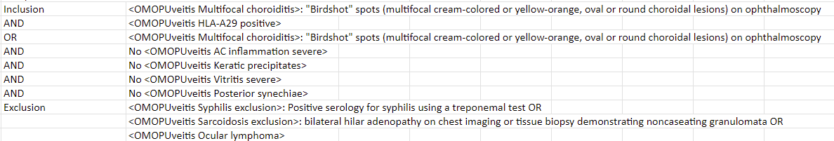
- Clinical description: Computable definition of SUN classification criteria for Birdshot chorioretinitis. This phenotype is of patients with a diagnosis or clinical findings of posterior uveitis consistent with birdshot chorioretinitis. This phenotype operationalizes the definition published by the SUN workgroup (PMID 33845003)
Criteria ([#’s 1, 2, and 3] OR # 4)
- Characteristic bilateral multifocal choroiditis on ophthalmoscopy
a. Multifocal cream-colored or yellow-orange, oval or round choroidal lesions (“birdshot spots”)
AND - Absent to mild anterior chamber inflammation
a. Absent to mild anterior chamber cells AND
b. No keratic precipitates AND
c. No posterior synechiae
AND - Absent to moderate vitritis
OR - Multifocal choroiditis with
a. Positive HLA-A29 test AND either (b. or c.)
b. Characteristic “birdshot” spots (multifocal cream-colored or yellow-orange, oval or round choroidal lesions) on ophthalmoscopy OR
c. Characteristic indocyanine green angiogram (multifocal hypofluorescent spots) without characteristic “birdshot” spots on ophthalmoscopy
Exclusions - Positive serologic test for syphilis using a treponemal test
- Evidence of sarcoidosis (either bilateral hilar adenopathy on chest imaging or tissue biopsy demonstrating non-caseating granulomata)*
- Evidence of intraocular lymphoma on diagnostic vitrectomy or tissue biopsy
-
*Logic description:
-
Recommended Study applications: target
-
Submitted cohort definition:: [ATLAS: Cohort Definitions (ohdsi.org) ](ATLAS: Cohort Definitions (ohdsi.org)